Scientific Image Gallery
Welcome to our Scientific Image Gallery. Here you can find real-life examples of cell images, mostly (but not only) from peripheral blood films, that illustrate typical morphologic characteristics pointing to specific conditions or disorders. This constitutes their diagnostic value.
Click on an image to enlarge it and display a short description.

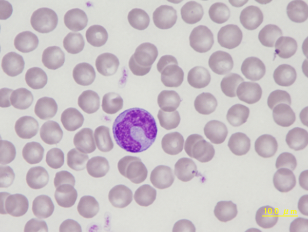
Monocyte with delicate nuclear chromatin and grey-blue cytoplasm. On close inspection, there are vacuoles in the cytoplasm which help to distinguish this monocyte from a metamyelocyte.
<p>Monocyte with delicate nuclear chromatin and grey-blue cytoplasm. On close inspection, there are vacuoles in the cytoplasm which help to distinguish this monocyte from a metamyelocyte.</p>

Canine monocytes show variably shaped nuclei with a clearly visible chromatin structure and occasionally faint, dust-like azurophilic granules evenly spread throughout the blue cytoplasm.
<p>Canine monocytes show variably shaped nuclei with a clearly visible chromatin structure and occasionally faint, dust-like azurophilic granules evenly spread throughout the blue cytoplasm. </p>

Erythroblast (nucleated red blood cell) showing a dark purple nucleus with condensed chromatin. The cytoplasm is polychromatic with the blue colour of ribosomes (RNA) mixed with the red colour of haemoglobin. The cell is slightly larger than a mature red blood cell. After enucleation this cell becomes a reticulocyte.
<p>Erythroblast (nucleated red blood cell) showing a dark purple nucleus with condensed chromatin. The cytoplasm is polychromatic with the blue colour of ribosomes (RNA) mixed with the red colour of haemoglobin. The cell is slightly larger than a mature red blood cell. After enucleation this cell becomes a reticulocyte.</p>

Supravital staining of a blood smear with new methylene blue showing three reticulocytes. Various reticulated structures including RNA-containing microsomes are stained.
<p>Supravital staining of a blood smear with new methylene blue showing three reticulocytes. Various reticulated structures including RNA-containing microsomes are stained. </p>

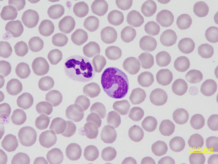
Typical segmented neutrophil on the left and a band neutrophil to the right. The band cell is immature and shows bluish cytoplasm, which changes to a lighter colour as the cell matures.
<p>Typical segmented neutrophil on the left and a band neutrophil to the right. The band cell is immature and shows bluish cytoplasm, which changes to a lighter colour as the cell matures. </p>

Segmented neutrophil with partly constricted nucleus. Monocyte with delicate nuclear chromatin and fine azurophilic granules in the cytoplasm. The shape of the nucleus in this monocyte is frequently seen in dogs.
<p>Segmented neutrophil with partly constricted nucleus. Monocyte with delicate nuclear chromatin and fine azurophilic granules in the cytoplasm. The shape of the nucleus in this monocyte is frequently seen in dogs.</p>

Blood smear of a 6-year-old male Pomeranian dog suffering from hepatic dysfunction associated with cholestasis and pancreatitis. Several spherocytes lacking the typical central pallor of canine red blood cells can be seen. In the centre, there is a band neutrophil.
<p>Blood smear of a 6-year-old male Pomeranian dog suffering from hepatic dysfunction associated with cholestasis and pancreatitis. Several spherocytes lacking the typical central pallor of canine red blood cells can be seen. In the centre, there is a band neutrophil. </p>

Transitional form of chronic lymphocytic leukaemia/prolymphocytic leukaemia with a white blood cell count of 250,000/μL. Prolymphocytes are indicated (->).
<p>Transitional form of chronic lymphocytic leukaemia/prolymphocytic leukaemia with a white blood cell count of 250,000/μL. Prolymphocytes are indicated (->).</p>

Chronic lymphocytic leukaemia with numerous prolymphocytes (->) (CLL/PL) in the peripheral blood (May-Grünwald-Giemsa stain).
<p>Chronic lymphocytic leukaemia with numerous prolymphocytes (->) (CLL/PL) in the peripheral blood (May-Grünwald-Giemsa stain). </p>