Scientific Image Gallery
Welcome to our Scientific Image Gallery. Here you can find real-life examples of cell images, mostly (but not only) from peripheral blood films, that illustrate typical morphologic characteristics pointing to specific conditions or disorders. This constitutes their diagnostic value.
Click on an image to enlarge it and display a short description.

An apparent Howell-Jolly body, caused by contaminated microscope oil, can be identified as an artefact by the fact that it moves independently over time.
<p>An apparent Howell-Jolly body, caused by contaminated microscope oil, can be identified as an artefact by the fact that it moves independently over time.</p>

Incidental detection of Borrelia recurrentis (->) in a patient initially suspected to suffer from malaria. Borrelia recurrentis is transmitted by lice and ticks and is the causative agent of relapsing fever. Like malaria, relapsing fever often is a travel-related disease that, after an incubation time of up to 2 weeks, leads to fever attacks.
<p>Incidental detection of Borrelia recurrentis (->) in a patient initially suspected to suffer from malaria. Borrelia recurrentis is transmitted by lice and ticks and is the causative agent of relapsing fever. Like malaria, relapsing fever often is a travel-related disease that, after an incubation time of up to 2 weeks, leads to fever attacks.</p>

In the peripheral blood (May-Grünwald-Giemsa stain) of this 75-year old woman with PV all three cell lineages are increased: haemoglobin concentration 16 g/dL, white blood cell count 15,000/µL, platelet count 980,000/µL. In functional iron deficiency red blood cells often are microcytic. The MCV is only 75 fL. The haematopoietic cells of the patient show a JAK2 mutation.
<p>In the peripheral blood (May-Grünwald-Giemsa stain) of this 75-year old woman with PV all three cell lineages are increased: haemoglobin concentration 16 g/dL, white blood cell count 15,000/µL, platelet count 980,000/µL. In functional iron deficiency red blood cells often are microcytic. The MCV is only 75 fL. The haematopoietic cells of the patient show a JAK2 mutation.</p>

Infectious mononucleosis is an acute condition caused by the Epstein-Barr virus (EBV). The disease is highly contagious and spreads via body secretions, especially saliva. The infection frequently goes unnoticed in children; however, mainly adolescents and young adults exhibit symptoms. Infected B lymphocytes induce a humoral (B cell) as well as a cellular (T cell) immune response which can be seen in an increased concentration of atypical lymphocytes in the blood film.
<p>Infectious mononucleosis is an acute condition caused by the Epstein-Barr virus (EBV). The disease is highly contagious and spreads via body secretions, especially saliva. The infection frequently goes unnoticed in children; however, mainly adolescents and young adults exhibit symptoms. Infected B lymphocytes induce a humoral (B cell) as well as a cellular (T cell) immune response which can be seen in an increased concentration of atypical lymphocytes in the blood film. </p>

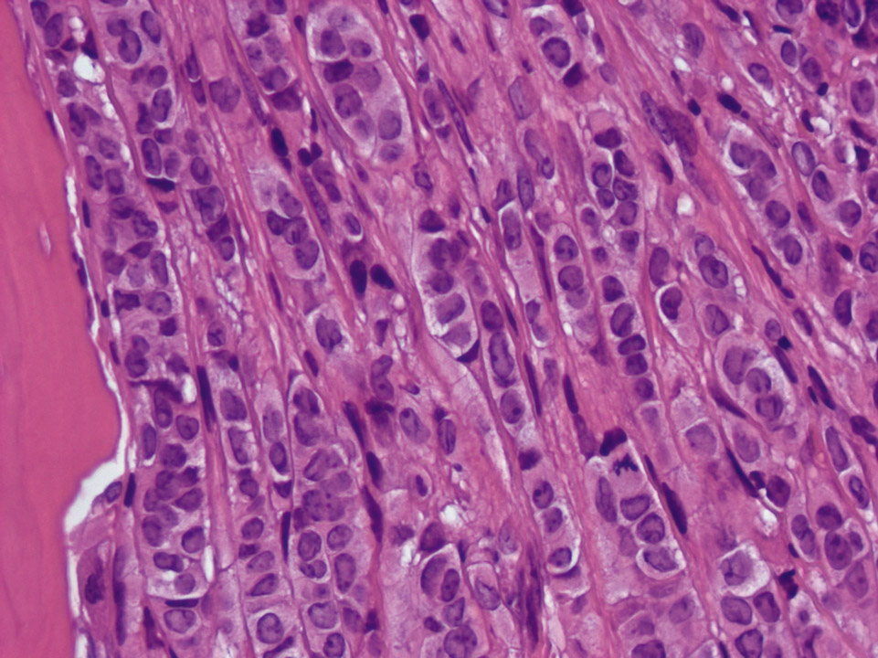
Bone marrow histology (haematoxylin eosin stain) of a patient with breast cancer showing an infiltration of the bone marrow by a lobular carcinoma of the breast.
<p>Bone marrow histology (haematoxylin eosin stain) of a patient with breast cancer showing an infiltration of the bone marrow by a lobular carcinoma of the breast.</p>

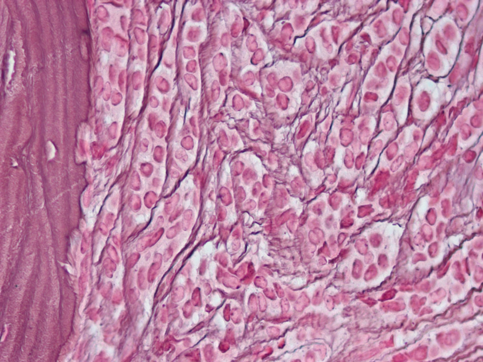
Bone marrow histology of a patient with breast cancer showing an infiltration of the bone marrow by a lobular carcinoma of the breast. The increased number of fibres is coloured black after Gomori staining.
<p>Bone marrow histology of a patient with breast cancer showing an infiltration of the bone marrow by a lobular carcinoma of the breast. The increased number of fibres is coloured black after Gomori staining.</p>

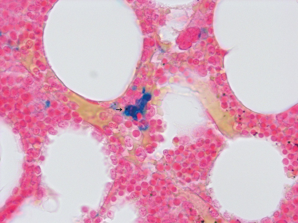
Due to an increased production of red blood cells in PV iron in the bone marrow gets 'used up', causing functional iron deficiency in the bone marrow. The detection of iron accumulation (->) in the bone marrow by Prussian Blue stain, as shown here, argues against PV.
<p>Due to an increased production of red blood cells in PV iron in the bone marrow gets 'used up', causing functional iron deficiency in the bone marrow. The detection of iron accumulation (->) in the bone marrow by Prussian Blue stain, as shown here, argues against PV.</p>

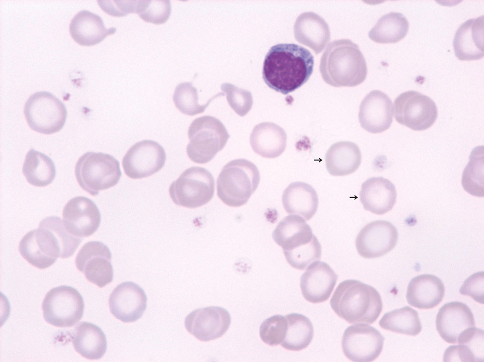
Patient with established severe iron deficiency anaemia (haemoglobin 5 g/dL). Well recognisable are anulocytes (->) and unusually small red blood cells – compared to a normal lymphocyte. The MCV was 53 fL.
<p>Patient with established severe iron deficiency anaemia (haemoglobin 5 g/dL). Well recognisable are anulocytes (->) and unusually small red blood cells – compared to a normal lymphocyte. The MCV was 53 fL.</p>