Scientific Image Gallery
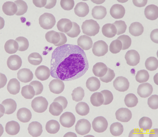
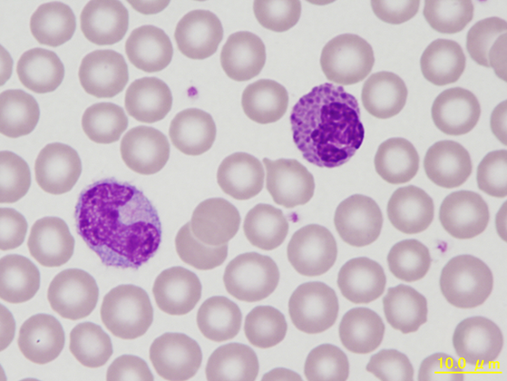
Welcome to our Scientific Image Gallery. Here you can find real-life examples of cell images, mostly (but not only) from peripheral blood films, that illustrate typical morphologic characteristics pointing to specific conditions or disorders. This constitutes their diagnostic value.
Click on an image to enlarge it and display a short description.
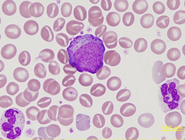
Neutrophilic myelocyte with many azurophilic granules in comparison to two mature, segmented neutrophils. Immature blood cells are an occasional finding in the peripheral blood of marmosets.
<p>Neutrophilic myelocyte with many azurophilic granules in comparison to two mature, segmented neutrophils. Immature blood cells are an occasional finding in the peripheral blood of marmosets.</p>
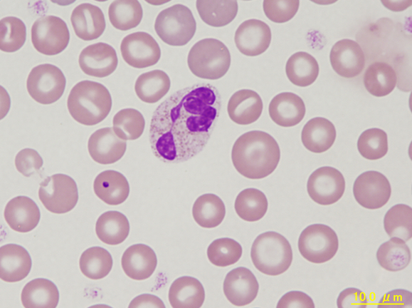
Marmoset blood smear showing a neutrophil with segmented nucleus and granules that appear eosinophilic. The red blood cell to the left contains a Howell-Jolly body, a common finding in the marmoset.
<p>Marmoset blood smear showing a neutrophil with segmented nucleus and granules that appear eosinophilic. The red blood cell to the left contains a Howell-Jolly body, a common finding in the marmoset.</p>
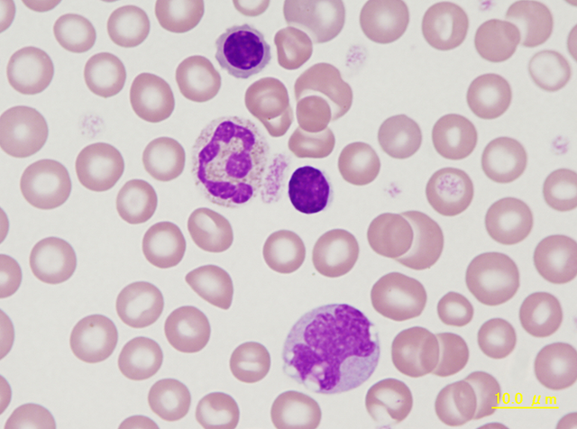
Typical neutrophil, lymphocyte (centre) and monocyte (bottom) of a marmoset. At the top there is an erythroblast showing condensed chromatin and a greater amount of slightly reddish cytoplasm reflecting haemoglobin synthesis. Giant platelet between the neutrophil and lymphocyte.
<p>Typical neutrophil, lymphocyte (centre) and monocyte (bottom) of a marmoset. At the top there is an erythroblast showing condensed chromatin and a greater amount of slightly reddish cytoplasm reflecting haemoglobin synthesis. Giant platelet between the neutrophil and lymphocyte.</p>
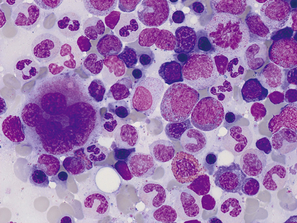
Normal bone marrow cytology. Some cells are always squeezed and damaged. These cells are called smudge cells. Smudge cells cannot be examined and must not be regarded as pathological (They have nothing to do with Gumprecht shadows in the peripheral blood smear). On the top right a mitosis is visible.
<p>Normal bone marrow cytology. Some cells are always squeezed and damaged. These cells are called smudge cells. Smudge cells cannot be examined and must not be regarded as pathological (They have nothing to do with Gumprecht shadows in the peripheral blood smear). On the top right a mitosis is visible. </p>
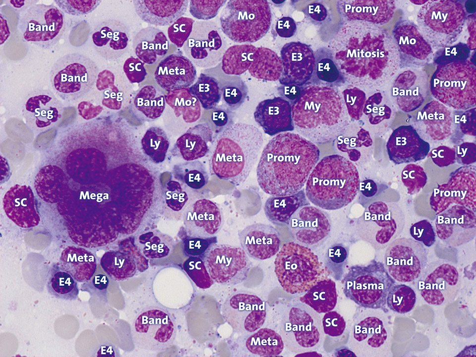
The abbreviations mean: Band: Band neutrophil, E3: Polychromatic erythroblast, E4: Orthochromatic erythroblast, Eo: Eosinophil granulocyte, Ly: Lymphocyte, Mega: Megakaryocyte, Meta: Metamyelocyte, Mo: Monocyte, My: Myelocyte, Plasma: Plasma cell, Promy: Promyelocyte, SC: Smudge cell, Seg: Segmented neutrophil, ?: No clear assignment possible.
<p>The abbreviations mean: Band: Band neutrophil, E3: Polychromatic erythroblast, E4: Orthochromatic erythroblast, Eo: Eosinophil granulocyte, Ly: Lymphocyte, Mega: Megakaryocyte, Meta: Metamyelocyte, Mo: Monocyte, My: Myelocyte, Plasma: Plasma cell, Promy: Promyelocyte, SC: Smudge cell, Seg: Segmented neutrophil, ?: No clear assignment possible.</p>
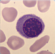
Cell description: 3 different stages of nucleated red blood cells are known: basophilic, polychromatic and orthochromatic
Size: around 10 µm
Nucleus: round with variable degree of chromatin condensation according to maturation, faint or absent nucleoli
Cytoplasm: bluish to pink (depending on maturation), no granules
Nucleus decreases in size as the cell matures
<p>Cell description: 3 different stages of nucleated red blood cells are known: basophilic, polychromatic and orthochromatic </p> <p>Size: around 10 µm </p> <p>Nucleus: round with variable degree of chromatin condensation according to maturation, faint or absent nucleoli </p> <p>Cytoplasm: bluish to pink (depending on maturation), no granules </p> <p>Nucleus decreases in size as the cell matures</p>
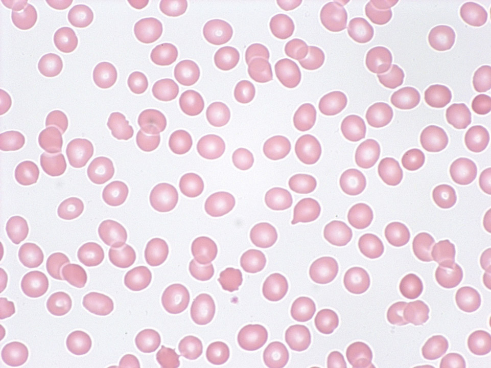
Pancytopenia (tricytopenia) in the peripheral blood (May-Grünwald-Giemsa stain) of a patient with severe aplastic anaemia: The automated cell count showed granulocytopenia (300/µL), mild lymphocytopenia (800/µL), anaemia (haemoglobin 9 g/dL), a reduced reticulocyte count (18,000/µL) and thrombocytopenia (12,000/µL).
<p>Pancytopenia (tricytopenia) in the peripheral blood (May-Grünwald-Giemsa stain) of a patient with severe aplastic anaemia: The automated cell count showed granulocytopenia (300/µL), mild lymphocytopenia (800/µL), anaemia (haemoglobin 9 g/dL), a reduced reticulocyte count (18,000/µL) and thrombocytopenia (12,000/µL).</p>
