Scientific Image Gallery
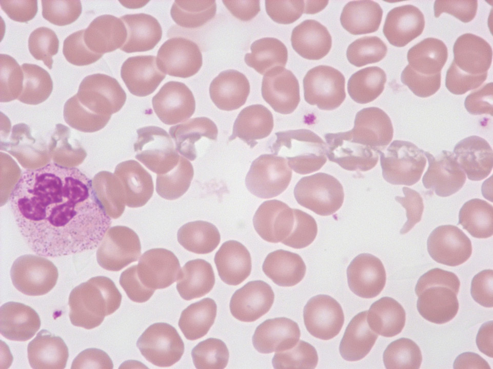
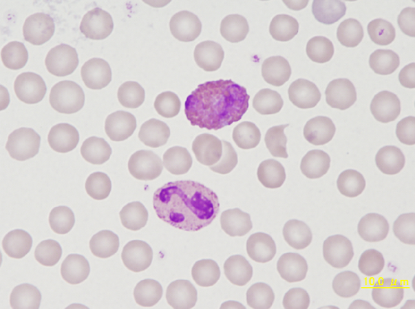
Welcome to our Scientific Image Gallery. Here you can find real-life examples of cell images, mostly (but not only) from peripheral blood films, that illustrate typical morphologic characteristics pointing to specific conditions or disorders. This constitutes their diagnostic value.
Click on an image to enlarge it and display a short description.
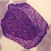
Cell description:
Size: 15-25 µm
Nucleus: oval with identifiable nucleoli and diffuse chromatin structure Cytoplasm: basophilic with visible golgi-zone and eye-catching azurophil granula (primary granulation)
<p>Cell description: </p> <p>Size: 15-25 µm </p> <p>Nucleus: oval with identifiable nucleoli and diffuse chromatin structure Cytoplasm: basophilic with visible golgi-zone and eye-catching azurophil granula (primary granulation)</p>
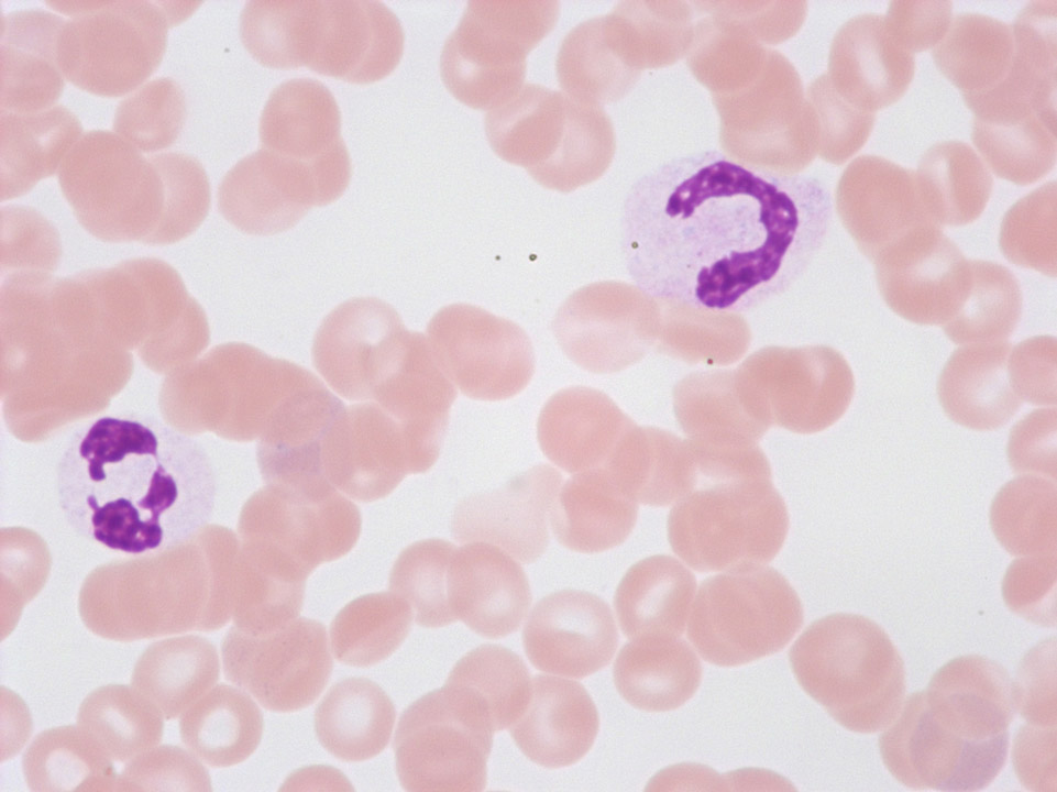
These so-called 'pseudo-Pelger's nuclear anomalies' with abnormal chromatin condensation and/or deficient nuclear segmentation are often found with viral infections, after chemotherapy, or with a myelodysplastic syndrome (MDS). Conversely, the true Pelger's nuclear anomaly is hereditary and without pathological significance.
<p>These so-called 'pseudo-Pelger's nuclear anomalies' with abnormal chromatin condensation and/or deficient nuclear segmentation are often found with viral infections, after chemotherapy, or with a myelodysplastic syndrome (MDS). Conversely, the true Pelger's nuclear anomaly is hereditary and without pathological significance.</p>
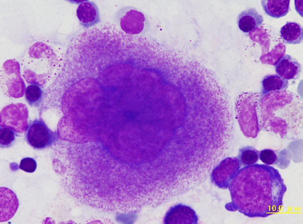
Bone marrow smear of a rabbit showing a megakaryocyte. The nucleus is lobulated and many azurophilic granules are found in the cytoplasm, suggesting maturity of the cell.
<p>Bone marrow smear of a rabbit showing a megakaryocyte. The nucleus is lobulated and many azurophilic granules are found in the cytoplasm, suggesting maturity of the cell. </p>
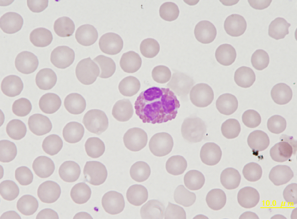
Eosinophil with large (approx. 1,5 µm) round and orange-reddish staining granules that are sharply defined and markedly larger than eosinophil granules in mice and rats.
<p>Eosinophil with large (approx. 1,5 µm) round and orange-reddish staining granules that are sharply defined and markedly larger than eosinophil granules in mice and rats.</p>
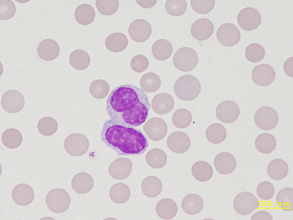
Two lymphocytes of a rabbit. The upper cell has a constricted nucleus which is more often seen with monocytes, but lacks the colour characteristic of monocyte cytoplasm.
<p>Two lymphocytes of a rabbit. The upper cell has a constricted nucleus which is more often seen with monocytes, but lacks the colour characteristic of monocyte cytoplasm.</p>

Monocyte on top, heterophil below. Similar to eosinophils, the granules of leporine neutrophils stain intensely red by May-Giemsa or Wright staining. Therefore, they are termed pseudoeosinophils or heterophils in the rabbit.
<p>Monocyte on top, heterophil below. Similar to eosinophils, the granules of leporine neutrophils stain intensely red by May-Giemsa or Wright staining. Therefore, they are termed pseudoeosinophils or heterophils in the rabbit.</p>
